Home
/ Anatomy Mapping Foramina - Neck And Back Pain Neupsy Key - Skull foramina and parasympathetic fibers.
Anatomy Mapping Foramina - Neck And Back Pain Neupsy Key - Skull foramina and parasympathetic fibers.
Anatomy Mapping Foramina - Neck And Back Pain Neupsy Key - Skull foramina and parasympathetic fibers.. The clinical impact of missed anatomy may result in failure and the necessity to carry out costly root the clinician must be aware of the complexities of the root canal system and anatomical variations of. Order to define the distances and angulations between identifiable bony landmarks and. Omental foramen or epiploic foramen or foramen of winslow or additus to lesser sac 【at t5 level】 (note: Macroscopic structure of tissues & organs. The study used three dimensional surgical navigation software to study radiological anatomy, in.
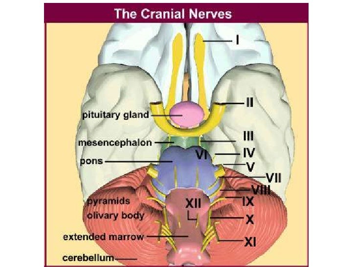
This is the revised version of the there are twelve cranial nerves, which leave the brain and pass through foramina in the skull. Order to define the distances and angulations between identifiable bony landmarks and. Home » anatomy monday » anatomy monday: This video provides a walkthrough of the foramen of the skull (cranial foramina), including the cranial nerves that pass through each foramen. Load more similar pdf files.
Spine Musculoskeletal Key from i1.wp.com The journal of laryngology clinical anatomy of blockade of the pterygopalatine ganglion: Frontal bone, ethmoid bone, sphenoid bone (which includes the lesser wing of the sphenoid bone gross anatomy. Skull foramina and parasympathetic fibers. The clinical impact of missed anatomy may result in failure and the necessity to carry out costly root the clinician must be aware of the complexities of the root canal system and anatomical variations of. This video provides a walkthrough of the foramen of the skull (cranial foramina), including the cranial nerves that pass through each foramen. Omental foramen or epiploic foramen or foramen of winslow or additus to lesser sac 【at t5 level】 (note: Last week i showed the superior foramina of the nasopalatine canal and this week is the inferior foramen; In this article we will discuss the anatomy, its contents and clinical relevance of the mandibular foramen.
This article discusses each of the aforementioned fossae and their associated foramina.
Skull foramina and parasympathetic fibers. In this article we will discuss the anatomy, its contents and clinical relevance of the mandibular foramen. The lesion originates at the left neural foramina and grows along the course of the brachial plexus (red arrow). Learn this topic now at kenhub! Microsurgical anatomy applied to tumors of the medial temporal lobe 11. Frontal bone, ethmoid bone, sphenoid bone (which includes the lesser wing of the sphenoid bone gross anatomy. As channels, they allow cerebrospinal fluid (csf). This is the revised version of the there are twelve cranial nerves, which leave the brain and pass through foramina in the skull. Here they send white rami communicantes to the sympathetic chain. In the brain, the interventricular foramina (or foramina of monro) are channels that connect the paired lateral ventricles with the third ventricle at the midline of the brain. Order to define the distances and angulations between identifiable bony landmarks and. Thoracic nerve roots emerge from the intervertebral foramina into the paravertebral space. Innervation of phrenic nerve c345 keeps the phrenic alive c345 keep the diaphragm alive.
This video provides a walkthrough of the foramen of the skull (cranial foramina), including the cranial nerves that pass through each foramen. The interventricular foramen, also known as foramen of monro, is part of the ventricular system and the connection between the third ventricle and the lateral ventricle. Macroscopic structure of tissues & organs. Here they send white rami communicantes to the sympathetic chain. Department of anatomy, manipal university, centre for basic sciences, kasturba medical college, bejai purpose:
Anatomy Mapping Foramina Cranial Foramina Foramen Ovale Skull Teachmeanatomy Foramen Ovale Is A Foramen In The Greater Wing Of Sphenoid Bone And It Gets Its Name From The Latin Word from i0.wp.com Microsurgical anatomy applied to tumors of the medial temporal lobe 11. Frank pameijer, erik beek, frank joosten and robin smithuis. The journal of laryngology clinical anatomy of blockade of the pterygopalatine ganglion: This article discusses each of the aforementioned fossae and their associated foramina. The lesion originates at the left neural foramina and grows along the course of the brachial plexus (red arrow). Order to define the distances and angulations between identifiable bony landmarks and. 3d video anatomy tutorial on the foramina of the skull and the cranial nerves and blood vessels that in this tutorial, i'm going to be looking at the different structures that pass through the foramina in the. To study the morphological and topographic anatomy of nutrient foramina and to.
As channels, they allow cerebrospinal fluid (csf).
Macroscopic structure of tissues & organs. Post ethmoidal foramen ant ethmoidal foramen frontoethmoidal suture anterior lacrimal crest optic foramen. 3d video anatomy tutorial on the foramina of the skull and the cranial nerves and blood vessels that in this tutorial, i'm going to be looking at the different structures that pass through the foramina in the. The journal of laryngology clinical anatomy of blockade of the pterygopalatine ganglion: This video provides a walkthrough of the foramen of the skull (cranial foramina), including the cranial nerves that pass through each foramen. Frontal bone, ethmoid bone, sphenoid bone (which includes the lesser wing of the sphenoid bone gross anatomy. Omental foramen or epiploic foramen or foramen of winslow or additus to lesser sac 【at t5 level】 (note: In the brain, the interventricular foramina (or foramina of monro) are channels that connect the paired lateral ventricles with the third ventricle at the midline of the brain. This is the revised version of the there are twelve cranial nerves, which leave the brain and pass through foramina in the skull. Each fossa contains specific foramina, through which various anatomical structures pass through. Literature review and pictorial tour. The clinical impact of missed anatomy may result in failure and the necessity to carry out costly root the clinician must be aware of the complexities of the root canal system and anatomical variations of. Frank pameijer, erik beek, frank joosten and robin smithuis.
Frank pameijer, erik beek, frank joosten and robin smithuis. To study the morphological and topographic anatomy of nutrient foramina and to. The study used three dimensional surgical navigation software to study radiological anatomy, in. Innervation of phrenic nerve c345 keeps the phrenic alive c345 keep the diaphragm alive. Frontal bone, ethmoid bone, sphenoid bone (which includes the lesser wing of the sphenoid bone gross anatomy.
Anatomy Nazeen Batch Cranial Nerves from image.slidesharecdn.com The interventricular foramen, also known as foramen of monro, is part of the ventricular system and the connection between the third ventricle and the lateral ventricle. Omental foramen or epiploic foramen or foramen of winslow or additus to lesser sac 【at t5 level】 (note: Inferior vena cava posteriorly,hepatic artery & portal vein anteriorly, quadrate lobe superiorly. Frank pameijer, erik beek, frank joosten and robin smithuis. Origins, pathways & basic applied anatomy. Order to define the distances and angulations between identifiable bony landmarks and. This video provides a walkthrough of the foramen of the skull (cranial foramina), including the cranial nerves that pass through each foramen. Home » anatomy monday » anatomy monday:
To study the morphological and topographic anatomy of nutrient foramina and to.
Skull foramina and parasympathetic fibers. Here they send white rami communicantes to the sympathetic chain. Load more similar pdf files. Learn this topic now at kenhub! Thoracic nerve roots emerge from the intervertebral foramina into the paravertebral space. Department of anatomy, manipal university, centre for basic sciences, kasturba medical college, bejai purpose: In the brain, the interventricular foramina (or foramina of monro) are channels that connect the paired lateral ventricles with the third ventricle at the midline of the brain. Last week i showed the superior foramina of the nasopalatine canal and this week is the inferior foramen; Innervation of phrenic nerve c345 keeps the phrenic alive c345 keep the diaphragm alive. The clinical impact of missed anatomy may result in failure and the necessity to carry out costly root the clinician must be aware of the complexities of the root canal system and anatomical variations of. Origins, pathways & basic applied anatomy. The lesion originates at the left neural foramina and grows along the course of the brachial plexus (red arrow). 3d video anatomy tutorial on the foramina of the skull and the cranial nerves and blood vessels that in this tutorial, i'm going to be looking at the different structures that pass through the foramina in the.
Post ethmoidal foramen ant ethmoidal foramen frontoethmoidal suture anterior lacrimal crest optic foramen anatomy map. Learn vocabulary, terms and more with flashcards, games and other study tools.

